SCIENCE
The Wonderful Winning Images From The Nikon Small World Photomicrography Competition 2019

The Nikon Small World Photomicrography Competition has celebrated the microscopic world for forty-five years and in the period, allowed scientists and enthusiasts to show off their artistry of scientific and stunning imagery captured through a microscope. Over 2000 amazing pieces of microphotography were submitted for the competition with passionate micro-photographers from almost 100 different countries. This year’s winning images were picked of a turtle embryo who took the first place out of 20 winning photos for their scientific know-how and technical wizardry.
The wonderfully colorful image of a turtle was captured by microscopy technician Teresa Zgoda and recent university graduate Teresa Kugler, work done with precision and skill.
“The embryo was about an inch and a quarter long, a little smaller than a walnut,” Zgoda told IFLScience.
Those other winning pictures include an alligator and a California two-spot octopus embryo, as well as a pregnant planktonic crustacean and mosquito larva, also a frozen drop of water was transformed into a charming piece, and male Mosquito, Vitamin C… and other a mesmerizing things. Scroll down below and open the next page to check at the rest of the top 20 photos, that made it to the final winner’s list.
#20th Place: Blood Vessels Of A Murine (Mouse) Heart Following Myocardial Infarction (Heart Attack)

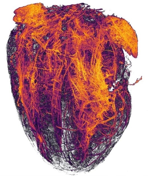
Simon Merz, Lea Bornemann & Sebastian Korste, University Hospital Essen, Institute for Experimental Immunology & Imaging, Essen, Nordrhein-Westfalen, Germany. Tissue Clearing, Light Sheet Fluorescence Microscopy, 2x (Objective Lens Magnification).
#19th Place: Octopus Bimaculoides Embryo

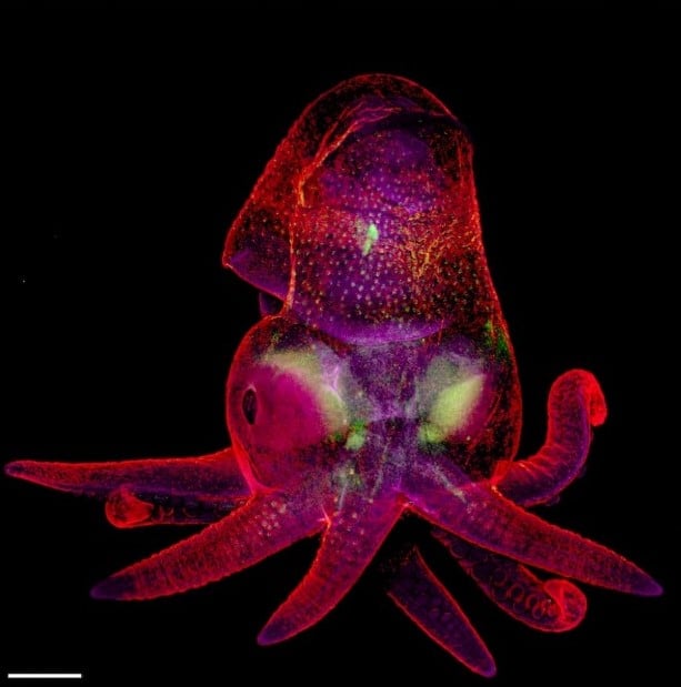
Martyna Lukoševičiūtė & Dr. Carrie Albertin, University of Oxford, Weatherall Institute of Molecular Medicine, Oxford, Oxfordshire, United Kingdom. Confocal, Image Stitching, 5x (Objective Lens Magnification).
#18th Place: Cristobalite Crystal Suspended In Its Quartz Mineral Host

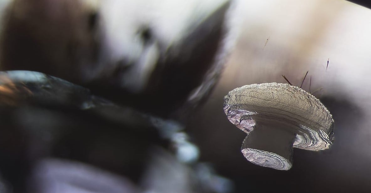
E. Billie Hughes, Lotus Gemology, Bangkok, Thailand. Darkfield, 40x (Objective Lens Magnification).
#17th Place: Vitamin C

Vitamin C. Karl Deckart (Eckental, Bavaria, Germany). Brightfield, Polarized Light. 4x (Objective Lens Magnification).
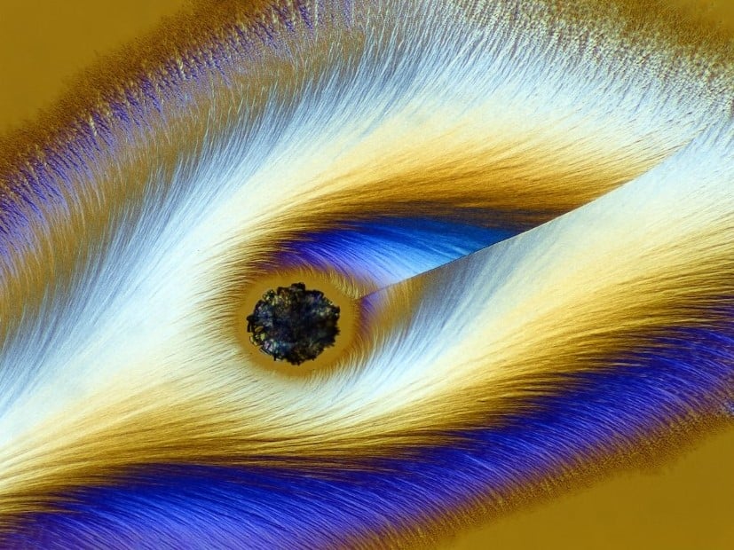
#16th Place: Housefly Compound Eye Pattern

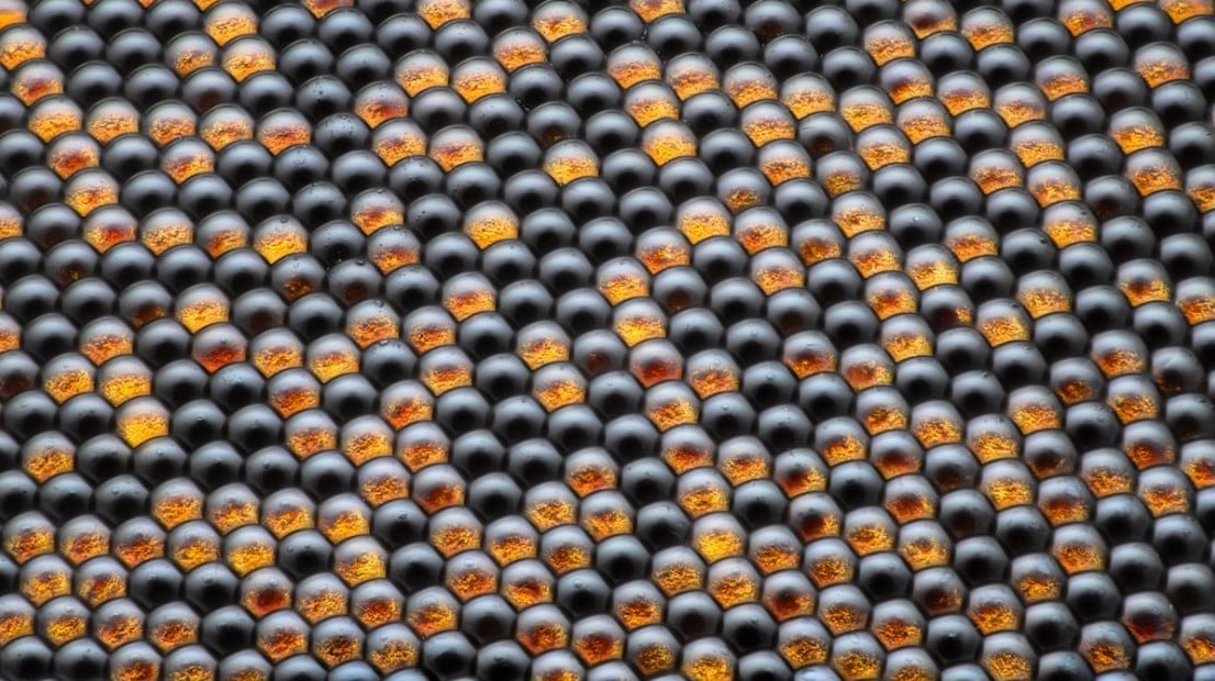
Dr. Razvan Cornel Constantin, Bucharest, Romania. Focus Stacking, Reflected Light, 50x (Objective Lens Magnification).
#15th Place: Pregnant Daphnia Magna (Small Planktonic Crustacean)


Marek Miś, Marek Miś Photography, Suwalki, Podlaskie, Poland. Modified Darkfield, Polarized Light, Image Stacking, 4x (Objective Lens Magnification).
#14th Place: Female Oxyopes Dumonti (Lynx) Spider

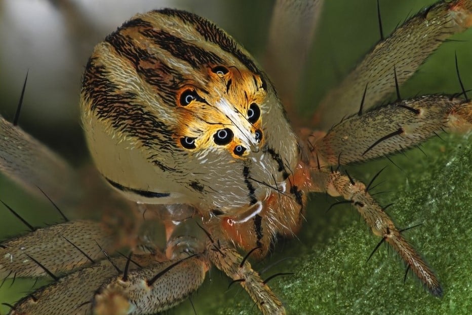
Antoine Franck, CIRAD – Agricultural Research for Development, Saint Pierre, Réunion. Focus Stacking, 1x (Objective Lens Magnification).
#13th Place: Cuprite (Mineral Composed Of Copper Oxide)

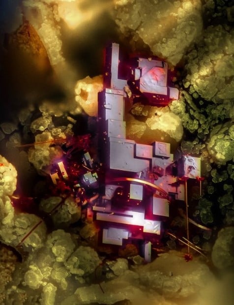
Dr. Emilio Carabajal Márquez, Madrid, Spain. Focus Stacking, 20x (Objective Lens Magnification).
#12th Place: Mosquito Larva

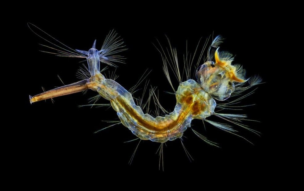
Anne Algar, Hounslow, Middlesex, United Kingdom. Darkfield, Polarizing Light, Image Stacking, 4x (Objective Lens Magnification).
#11th Place: A Pair Of Ovaries From An Adult Drosophila Female Stained For F-Actin (Yellow) And Nuclei (Green); Follicle Cells Are Marked By Gfp (Magenta)

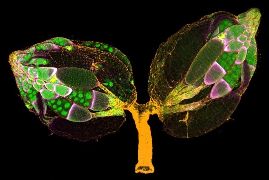
Dr. Yujun Chen & Dr. Jocelyn McDonald, Kansas State University, Department of Biology, Manhattan, Kansas, USA. Confocal, 10x (Objective Lens Magnification).
#10th Place: BPAE Cells In Telophase Stage Of Mitosis

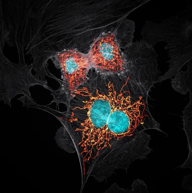
Jason M. Kirk, Baylor College of Medicine, Optical Imaging & Vital Microscopy Core, Houston, Texas, USA. Confocal with Enhanced Resolution, 63x (Objective Lens Magnification).
#9th Place: Tulip Bud Cross Section

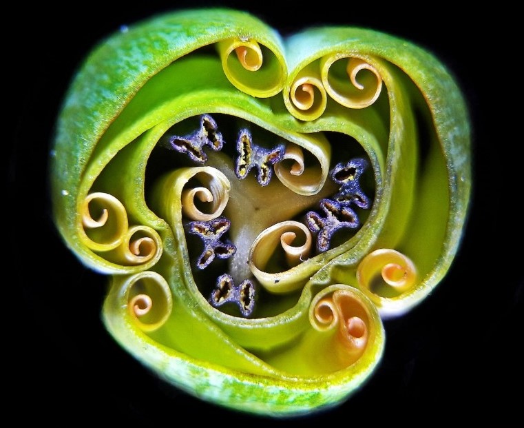
Andrei Savitsky, Cherkassy, Ukraine. Reflected Light, 1x (Objective Lens Magnification).
#8th Place: Frozen Water Droplet

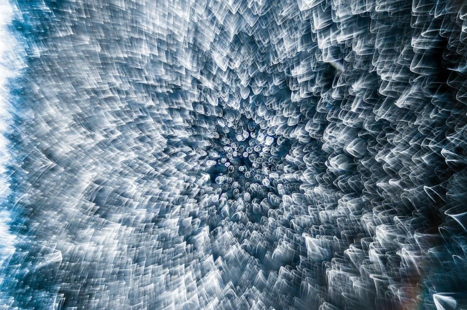
Garzon Christian, Quintin, Cotes-d’Armor, France. Incident Light, 8x (Objective Lens Magnification).
#7th Place: Chinese Red Carnation Stamen

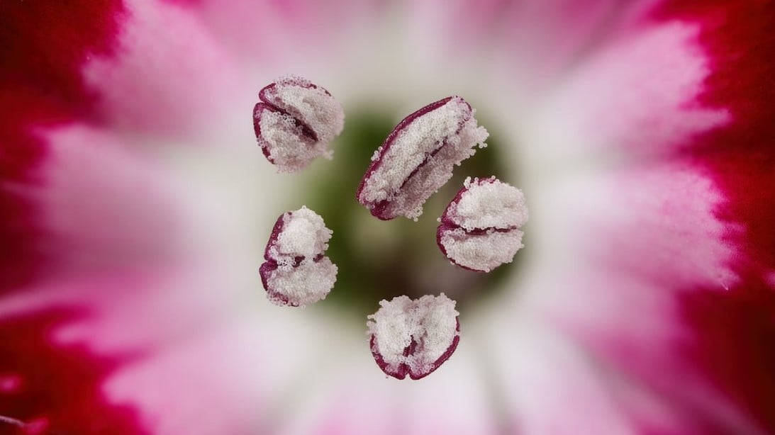
Dr. Guillermo López, Alicante, Spain. Focus Stacking, 3x (Objective Lens Magnification).
#6th Place: Small White Hair Spider

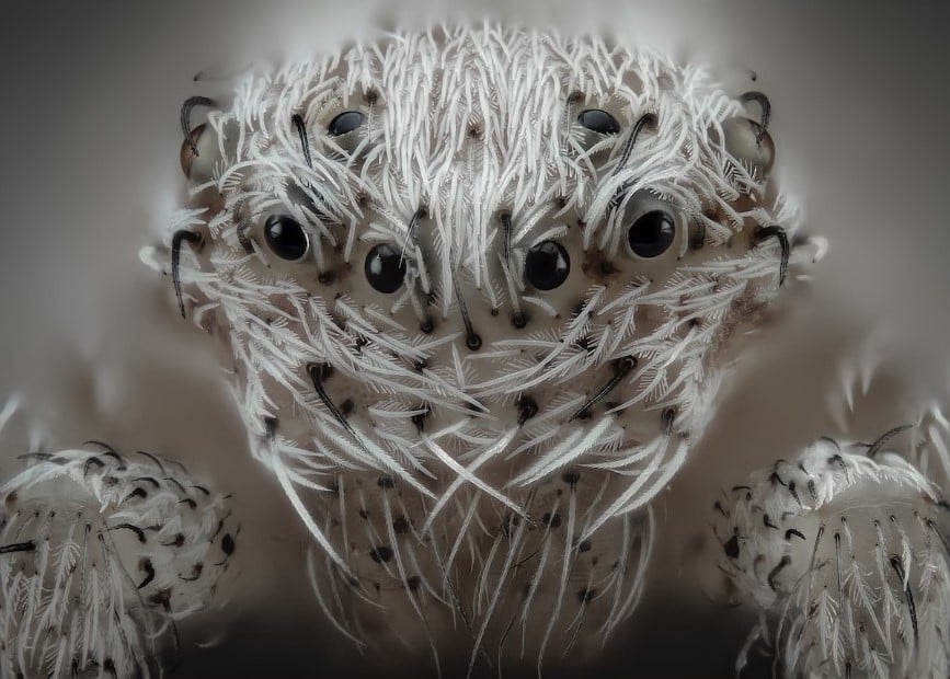
Javier Rupérez, Almáchar, Málaga, Spain. Reflected Light, Image Stacking, 20x (Objective Lens Magnification).
#5th Place: Snowflake

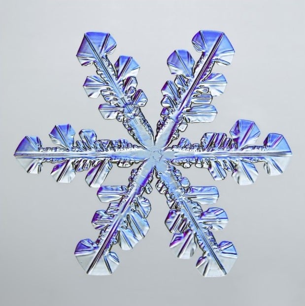
Caleb Foster, Caleb Foster Photography, Jericho, Vermont, USA. Transmitted Light, 4x (Objective Lens Magnification).
#4th Place: Male Mosquito

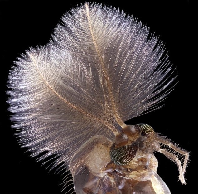
Jan Rosenboom, Universität Rostock, Rostock, Mecklenburg Vorpommern, Germany. Focus Stacking, 6.3x (Objective Lens Magnification).
#3rd Place: Alligator Embryo Developing Nerves And Skeleton

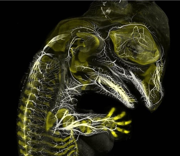
Daniel Smith Paredes & Dr. Bhart-Anjan S. Bhullar, Yale University, Department of Geology and Geophysics, New Haven, Connecticut, USA. Immunofluorescence, 10x (Objective Lens Magnification).
#2nd Place: Depth-Color Coded Projections Of Three Stentors (Single-Cell Freshwater Protozoans)

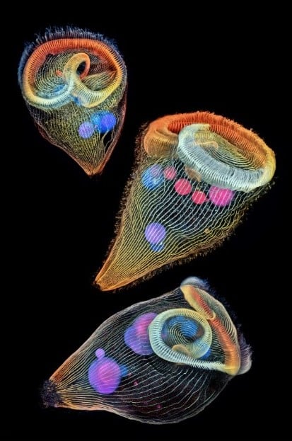
Dr. Igor Siwanowicz, Howard Hughes Medical Institute (HHMI), Janelia Research Campus, Ashburn, Virginia, USA. Confocal, 40x (Objective Lens Magnification).
#1st Place: Fluorescent Turtle Embryo

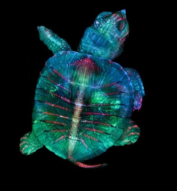
Teresa Zgoda & Teresa Kugler, Campbell Hall, New York, USA. Stereomicroscopy, Fluorescence, 5x (Objective Lens Magnification).